Could Ultra-Low Dose CT Scan Do The Trick?
How can ultra-low dose CT scans compare to standard CTs when creating 3D models for preoperative planning and surgery?
The number of people with knee disorders is steadily increasing in line with the mean age of the population. Osteoarthritis is a common cause of knee impairment, burdening affected people and society significantly. Knee arthroplasty, including uni-compartmental and total knee arthroplasty (UKA and TKA), is used to treat end-stage knee osteoarthritis. According to estimations, the number of knee arthroplasties performed in the United States among adults aged 65 increased by 162% between 1991 and 2010, with a 600% increase expected by 2030.
3D printing technology (3DP) has recently progressed by leaps and bounds. From using 3DP for aortic repairs to micromanaging plastic and neurosurgical cases to planning even the simplest outpatient surgeries, it is almost becoming an integral part of a surgical unit.
This technique is mainly utilized in orthopedics for pre-operative morphological design, intraoperative tissue repair, and major bone defect reconstruction. Using 3DP models and images during surgery has allowed for the resolution of complicated issues. Based on CT data, 3DP models give the surgeon a clearer anatomical perspective in full 3D, facilitating pre-operative digital planning of osteotomy. This strategy has shown incredible benefits in pre-operative planning, intraoperative precision, as well as reducing post-operative problems. Likewise, osteotomies performed using 3D-printed patient-specific guides appear to be a feasible therapy option for upper or lower limb malunion1.
The advantages of using 3DP become even more profound when operating on bone tumors. Because of the long-term use of the post-operative joint function, young tumor patients require near-perfect limb salvage surgery. The essential elements in determining the effect of the surgery are accurate and full excision and repair of joint function. Surgical guides combined with computed tomography (CT) three-dimensional reconstruction technology can considerably enhance osteotomy accuracy, reduce surgical complications, and improve joint reconstruction stability2.
Despite being an integral part of a hospital setting, CT scans come with the disadvantage of radiation exposure. Radiation exposure can potentially be the cause of major diseases. Although CT imaging investigations account for only 11% of radiologic operations, they account for over 70% of the total effective dosage.
The focus in CT imaging has been on lowering tube voltage and current. Recent research, however, has focused on ways to increase image quality using new algorithms. The lowering of voltage and current minimizes the dosage of radiation exposure directly. However, such a decrease unavoidably increases noise and reduces diagnostic accuracy. With the advancement of follow-up computer technology in recent years, iterative algorithms have been used in clinical settings to improve picture quality, particularly in low-radiation CT scanning. 3DP models using these algorithms have provided more than adequate data and far less radiation exposure. These models had a better picture quality and met clinical diagnostic objectives3.
One might think, how different the radiation exposure can be? The patient still gets the radiation. Right? Yes, it is right that the person would still be exposed to radiation, but the amount of radiation is DRASTICALLY reduced. The ultra-low dose CT group received 1.1 uSv of radiation, which is 33 times lower than chest X-rays and two times lower than hands and feet X-rays.
Stating the obvious benefit of low-dose exposure, various studies have also shown that models generated using ultra-low dose CT (ULD-CT) have not lagged while planning the operative management of the patient as compared with the low or normal-dose CT scanning. An article published in 2021 showed that the authors employed a 3-point assessment approach to analyze the 3D printed model’s definition and the operation’s direction. Three points were assigned when the 3D printed model’s smooth surface had no negative impact on surgical scheme planning, pre-operative operational practice, or intraoperative auxiliary operation; two points were rewarded when the 3DP model was slightly rough but had no negative impact on surgical planning and practice. One point was given only when the 3DP model had a rough surface that significantly impacted surgical scheme planning and pre-operative simulation. The study showed that there was a 97% of reduction in radiation exposure with little to no difference in subjective scoring of the 3D printing model4.
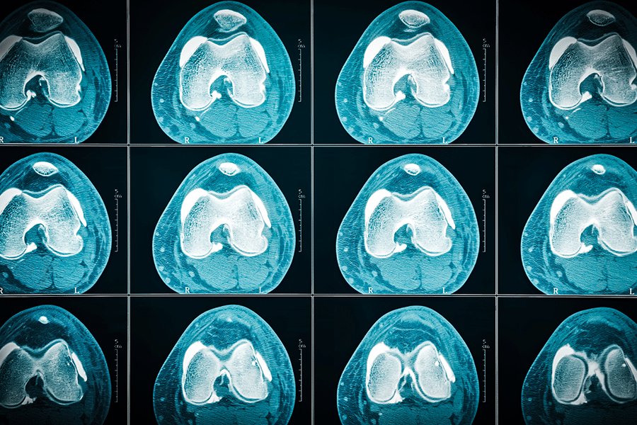
Another research published in 2022 compared the results of both regular and ULD-CT scans. A regular and ultra-low-dose CT scan was performed on the same subject. A 3D printed model was created with uniformly thick and spaced picture layers (0.5 mm each). Two 3D-printed knee joints were compared for their objective and perceptual differences. The researchers found no statistically significant difference between standard-dose and ultra-low-dose CT scans regarding femur, tibia, or patella size. In terms of clinical 3DP, the model’s surface was sufficiently smooth and fulfilled the requirements to aid in pre-operative planning and surgery.
The benefits of 3DP are not limited to the pre-operative surgical planning studies have shown that by using a 3D guide template, surgeons are now able to remove less bone and implant knee prostheses with more precision, saving time and reducing the risk of complications like, neurovascular damage from poorly placed screws making this all the more important to apply in daily practices of TKA5.
In conclusion, the present research indicates the viability of the 3D knee model utilizing the ULD-CT scan for pre-operative planning and TKA simulation. Ultra-low-dose imaging procedure for the knee lowers radiation exposure while retaining picture quality. ULD-CT met the requirements for 3DP model creation, internal fixation model selection, and simulated surgery. This approach is, therefore, suitable for application in clinical settings.
References:
1. Hoekstra, Harm, et al. “Corrective limb osteotomy using patient specific 3D-printed guides: a technical note.” Injury 47.10 (2016): 2375-2380.
2. Wang, Fengping, et al. “The application of 3D printed surgical guides in resection and reconstruction of malignant bone tumor.” Oncology Letters 14.4 (2017): 4581-4584.
3. Grosser, Oliver S., et al. “Iterative CT reconstruction in abdominal low-dose CT used for hybrid SPECT-CT applications: effect on image quality, image noise, detectability, and reader’s confidence.” Acta radiologica open 8.6 (2019): 2058460119856266.
4. Xiao, Mengqiang, et al. “Application of ultra-low-dose CT in 3D printing of distal radial fractures.” European Journal of Radiology 135 (2021): 109488.5. Ma, Limin, et al. “3D printed personalised titanium plates improve clinical outcome in microwave ablation of bone tumors around the knee.” Scientific reports 7.1 (2017): 1-10.
