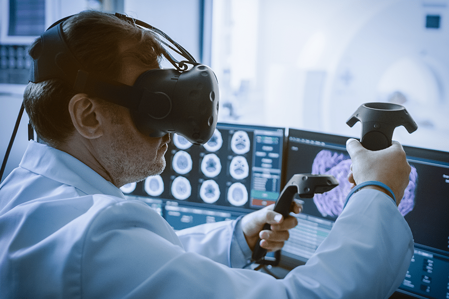
Virtual Reality Proves Useful For 3D Surgical Planning of Liver Surgery
A team of medical experts have developed a virtual reality application for viewing 3D models of the liver for use in surgical planning. 3D models are nothing new to the medical field. Complex surgeries often use 3D imagery to gain a more complete understanding of the anatomy or surgery in question. One of the primary issues that health providers face is that most 3D imagery is still viewed on a 2D computer screen. While this is still much more useful than examining a set of radiographs (x-rays), the spatial awareness and sense of depth and size can easily be lost due to the 2D nature of the display. This is where the authors of the study referenced in this post set out to improve the experience of viewing 3D models.
Virtual reality headsets are built to display 3D images. The popularity of VR has grown substantially in recent years, primarily due to the gaming industry. With a wide variety of headset models available to the general public, all with their own parameters, requirements, and prices, virtual reality has a relatively low cost of entry, especially when compared to most medical equipment. Most VR headsets are very similar in practice, requiring computing power to display and run the software, as well as a headset and set of controllers for interacting with the software. This makes it fairly easy for users to understand and use multiple different headsets without requiring a significant re-learning curve.
The researchers take an identical approach to 3D model generation as Kinomatic does with their joint replacements. It begins with a CT or MRI scan. Those images are then brought into another software which can take the CT or MRI data and convert it into three dimensional geometry. Everything is perfectly accurate to scale as it exists within the patient undergoing treatment, as it is a 1:1 digital recreation of their anatomy. They then separate the various aspects of the liver (parenchyma, tumors, hepatic veins, portal vein, and hepatic artery) as separate files. These files are exported as stereolithography (STL) files, which is the most common type of file for 3D printing. Whereas some researchers would go ahead and print these models for examination in a physical 3D form, this requires additional time and cost. Instead, they are able to upload the STL file directly to their application for near instant availability in the VR headset.
The researcher’s application makes it possible for the user to manipulate (rotate, zoom, reposition) the model in real time. They also allow the surgeon to change the opacity of the model from fully transparent to fully opaque. They also allow the user to draw potential resection lines on the model, and all of these features can be used in a single or multi user mode. They had five highly experienced surgeons evaluate the application on a usability scale out of 100% (0–50%: not acceptable, 51–68%: poor, 68%: OK; 69–80%: good, 81–100%: excellent). None of the surgeons had any previous experience using any virtual reality system. Despite this, the five surgeons scored an average of 76.6% justifying the ease of use of the VR application and all of them emphasized the potential for future utilization.
What the referenced article does a great job at is highlighting the difference between viewing 3D models on 2D screen versus in a fully interactive 3D, virtual reality application. They note that VR technology solves nearly all of the disadvantages of viewing 3D models on a 2D screen, while delivering nearly all of the advantages of actually 3D printing the models, thus saving time, money, and providing a better experience than what’s currently being done.
Virtual reality does have a few drawbacks. The ability to use this technology intraoperatively is certainly something that could be possible in the future, but because of the complete lack of sight, is not a viable option. When using the VR application to manipulate the models there is no haptic feedback, thus the feeling of actually holding or cutting anything in the virtual reality realm has yet to be accurately reproduced.
The important thing that Kinomatic, as well as the researchers mentioned in this post, have found is that the utility of VR in a surgical planning sense is an immense step forward from conventional methods of surgical planning. 3D models truly come to life when viewing them through the headset, and the learning curve is not as steep as many think. By providing surgeons with a new tool to view their patient anatomy in a way that most closely represents the real-life counterpart, the surgeon is able to assess the patient’s issue from a much more informed position than before. It is Kinomatic’s belief that virtual reality for surgical planning will only continue to advance and will soon become the new standard for viewing patient treatment plans.
To learn more about Kinomatic’s Virtual Reality Custom Surgical Planning, click here.
References:
Boedecker, C., Huettl, F., Saalfeld, P. et al. Using virtual 3D-models in surgical planning: workflow of an immersive virtual reality application in liver surgery. Langenbecks Arch Surg 406, 911–915 (2021). https://doi.org/10.1007/s00423-021-02127-7
